Introduction
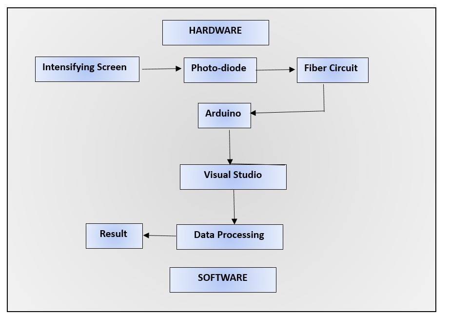
X-rays machines are playing important role in today’s medical world. The invention of X-rays made easy diagnosis of the problem by their imaging technique. The x-rays are allowed to pass through the target and then diffracted after hitting the object. The pattern obtained after diffraction is used to study the case. Initially this technology was used in orthopedics but later, developed its importance in every field. X-ray machines are essential medical instruments. However, if not used properly, they might be hazardous. The hazards include rashes, skin burn and other skin allergies. These hazards came under consideration when many scientists went through the same problem, which then was found to happen due to long term exposure to x-rays. If the voltage and exposure time is not controlled efficiently then it may result an explosion also. After going through all these serious hazards a problem arose, how to efficiently work with X-rays, as it was every useful inventions and it was a necessary tool in many fields such as in field of medicine so to ban it was not a solution, so scientists became to work on to devise a technique to work with X-rays efficiently. They created a unique tool to keep them precise and safe which was Indirect Measure Voltage and Exposure Time Device for Medical X-ray. There are two components to this device: intelligent software and hardware. With some smart hardware, X-ray energy is converted to light and subsequently to power. This power is properly handled before being transferred to Arduino. Like a smart detective, Arduino smells the duration of X-ray usage as well as a crucial aspect of the X-ray apparatus. The outcomes are then displayed in a Visual Studio. Thus, the work contributes to the safety and accuracy of X-ray machines, which eventually benefits patients.
An X-ray machine is a medical imaging gadget that employs X-rays to make pictures of the interior of the body. X-rays are a shape of electromagnetic radiation, and when coordinated through the body, they are refracted by distinctive tissues. This diffracted ray’s pattern makes a picture of the inner structures, permitting healthcare experts to analyze and screen different therapeutic conditions.
The fundamental working rule of an X-ray machine includes the era of X-rays, their controlled conveyance through the body, and the detection of the transmitted X-rays to make a picture. The X-ray machine contains an X-ray tube, which is the essential component capable for creating X-rays. Inside the X-ray tube, high-energy electrons are produced and quickened towards a metal target (more often than not tungsten). When these high-energy electrons strike the target, X-rays are delivered.
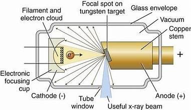
The interaction between the high-energy electrons and the metal target causes the inner-shell electrons of the target molecules to be catapulted. Electrons from external shells at that point fill these opening, and amid this prepare, X-rays are transmitted. The X-ray machine is prepared with a collimator, a gadget that shapes and limits the X-ray pillar.
The collimator permits the administrator to control the estimate and shape of the X-ray bar to center it on the particular zone of the body being inspected.
The persistent is situated between the X-ray tube and a finder (such as a film or a computerized sensor).The X-ray bar is coordinated through the patient’s body. As the X-rays pass through the body, they are retained to shifting degrees by distinctive tissues, depending on their thickness.
The X-rays that pass through the body make a shadow-like picture on the finder. Thick structures like bones assimilate more X-rays and show up as lighter ranges on the picture, whereas gentler tissues permit more X-rays to pass through and show up dark. In conventional X-ray frameworks, a film cassette captures the X-ray picture. The film is at that point created to form an unmistakable picture. In computerized X-ray frameworks, a computerized locator captures the X-ray picture electronically. This advanced data can be handled and shown on a computer screen.
The coming about X-ray picture is translated by a radiologist or a healthcare proficient. Anomalies, wounds, or other therapeutic conditions can be distinguished based on the appearance of structures within the X-ray.
Literature Overview
X-rays were discovered in 1869, when scientists were studying cathode rays. It was assumed that these rays were generated by the ionization of free electrons of the tube with the residual air at high voltage. Many scientists also discovered these rays but were not able to classify what actually it is. A Stanford university professor observed x rays while working on his technique of generation of electronic photograph. After this thing a concept was aired “Without Lens or Light, Photographs Taken with Plate and Object in Darkness “.
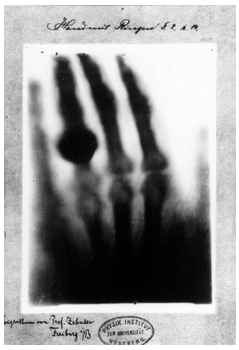
Rontgen was the first man to enlighten the use of x-rays for medical purposes. He was working on cathode and crook tubes when he accidently saw the x-rays. He flashed the x-rays for the medical purposes by creating the stricture of his wife’s hand.
After Rontgen’s this work, scientists started vast researches in x-rays for medical purposes and with passing time more challenges were faced which resulted in more accuracy and improvements. After this many tested X-rays on insects and other animals and concluded that X-rays not only photo the organ but it also disturbs the living function. Since X-rays were used in laborites and surgeries in medical uses.
Since x-rays were the new invention of that era so scientists were working devotedly in their research and harms were unknown. It was reported that many scientists faced burns, scars, hair fall and other skin allergies. At first it was a mystery but was known that it is due to the long term exposure to the X-rays. So precautionary measures were now the point of study.
Since many of hinders and barrier were discovered in X-rays work so scientists started their work in filed to improve the technique and minimize the hazards.
The first typical X-ray machine consisted of a Rumchoff coil connected to the cold cathode parallel to the vacuum tube. When the coil was heated the air gap between the cathode and the tube was ionized resulted in emission of rays. The drawback of this model was that it was high energy model and not reliable.
After this thermo-diode was invented which was connects to the hot cathode. But this was although better than previous but not reliable. In order to protect scientists from long-term exposure to X-rays it was decided to conceal the setup in a box. The rays will be produced inside the box and diffracted to the target. In this idea it was proposed to create a machine which generates x-rays inside a box and emitted to the target without being diffracted in order to prevent radiologists from long term exposure. This idea reduced the hazards to an extent but the problem to control the x-rays was still there. After so many researches and experiments an idea to control the exposure of x-rays externally was deduced by providing indirect voltage for x-rays emission and measuring the exposure externally which will be described in this research paper.
Methodology
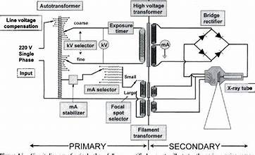
The X-ray circuit is structured into three distinct sections: the low-voltage circuit, filament circuit, and high-voltage circuit. Each section plays a crucial role in the overall functionality of the X-ray system.

The electrical potential difference between the X-ray tube’s cathode and anode is referred to as voltage in the context of X-ray machines. To create the electron flow necessary for the creation of X-rays, this potential difference is essential. A high-voltage generator is used to provide the high voltage required for medical X-ray imaging. Usually, an autotransformer, a rectifier, and an X-ray tube make up this generator. While the rectifier transforms AC electricity into DC, the autotransformer permits voltage modification. In X-ray imaging, kVp is an important parameter. It influences picture contrast and establishes the energy of X-ray photons. A precise choice of kVp is required for a correct diagnosis.
To measure the voltage output from the high-voltage generator indirectly, voltmeters or oscilloscopes are used. This non-invasive assessment makes sure the machine is working as efficiently as it should. To ensure accuracy, the voltage measurement instruments must be calibrated on a regular basis. To guarantee constant and secure voltage levels, quality control procedures are carried out, such as simulating patient exposure with a phantom.
The low-voltage circuit is the primary part of the device. It consists of a step down transformer which lowers the input voltage to the required value. This voltage is used to power up the parts of the device such as kvp meter, exposure time device and other small components. A low current is provided to warm the X-beam tube fiber, working with thermionic outflow of electrons.
The high-voltage circuit is a step up transformer fitting which is capable of emitting the x-rays. The low voltage coming from low voltage circuit is stepped up and powers up the tube to emit x-rays. The amount of x-rays to be emitted is controlled by indirectly controlling the voltage provided to the high voltage circuit as shown in Figure 7.
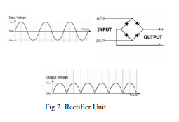
Because of full-wave correction, the beat x-beam yield happens at a recurrence of 100 times each second or 1 heartbeat for every 10 milliseconds (0.01 seconds). This data is crucial for computing the openness season of X-beams in the ensuing phases of the exploration.
A flat surface coated with fluorescent crystals (phosphors) that emit light (520-590 nm) when exposed to X-rays. A semiconductor device converting light into an electric current, with a sensitivity range between 350 to 820 nm and a maximum sensitivity wavelength of 550 nm. Utilizes a notch filter to reduce 50 Hz noise from electrical machines, preserving the X-ray signal at 100 Hz. Reads input from the photodiode’s pin and connects to a computer for signal reception and data processing, using the analog pin to receive signals.
The software component employs the Visual Studio program for receiving signals from Arduino and processing data. The Windows Form application on Visual Studio creates a user interface to display and process data received from the Arduino.

Conclusion
From this study we came to know how invention of x-rays has modernized the medical field. We came to know the technique of X-rays emission and the hazards that were cause during x-ray studies and the techniques discovered to overcome those hazards, among which the most reliable was indirect voltage measure and exposure time device. In this technique the voltage is applied separately to emit x-rays which are then controlled by varying the intensity of rays by varying the voltage and the exposure time of x-rays is set which, means to that particular extent x-rays will be emitted and after that the x-rays will be stopped generated automatically.
Future Work
By testing the given model 10% of error was calculated which is considered to be a precise but further improvements can be made to minimize this error for more accuracy. This can be accomplices by relating the kvp and exposure time in a manner that the delay observed in previous setting could be adjusted to have minimum gap.
Secondly long term exposure to x-rays is still harmful which still a problem is for radiologist. It suggested to work on x-rays. We should discover an element which provides fine x-rays but less hazardous or to emit x-rays through a source which directly hit the x-rays to the target without dispersing in the surrounding.
In the subject of X-rays, a number of improvements and possible future developments were being investigated. The following are some possible avenues for X-ray technology in the future:
Contrast Phase-Image Analysis: When compared to traditional X-ray imaging, this method offers superior contrast in soft tissues, making it possible to see structures like muscles and ligaments more clearly.
X-ray imaging with dual energy: This technology improves the ability to distinguish between various materials in the body by utilizing two distinct X-ray energies, hence providing improved diagnostic capabilities.
Reduced Radiation Dose: To lessen the possible hazards connected with X-ray exposure, researchers are still investigating ways to obtain high-quality photos at lower radiation doses.
Cone Beam CT: This technique produces three-dimensional pictures, which are very helpful for orthopedics and dentistry.
A 3D reconstruction of the region under examination is produced by taking several X-ray pictures from various angles, much like CT scans do.X-ray fluorescence imaging: This method may be used to identify and see certain components inside tissues, giving details on their concentration and distribution.
Automated Image Analysis: To help radiologists analyze X-ray images more accurately and efficiently, AI systems are being created.
Predictive modeling: Using patient data and X-ray results, AI may be used to forecast the likelihood of developing a disease.
X-ray devices that are portable and point-of-care: Miniaturization: Aims are being made to create more compact and transportable X-ray equipment for usage in emergency scenarios, point-of-care settings, and resource-constrained areas.
Applications in Therapy: With less harm to healthy tissues, micro beam radiation therapy is an experimental technology that uses narrow, high-dose X-ray beams for possible cancer treatment.
High-Energy X-ray Imaging: In areas like fabric science and mechanical testing, higher vitality X-rays are utilized to enter thick materials for superior review and examination.
Multimodal Imaging: Combining Imaging Modalities: Joining X-ray imaging with other imaging modalities such as MRI or PET can give comprehensive demonstrative data.
Authors:
- WALEED HANIF
- ALI HASSAN DAYAL